S-Shearwave Imaging™
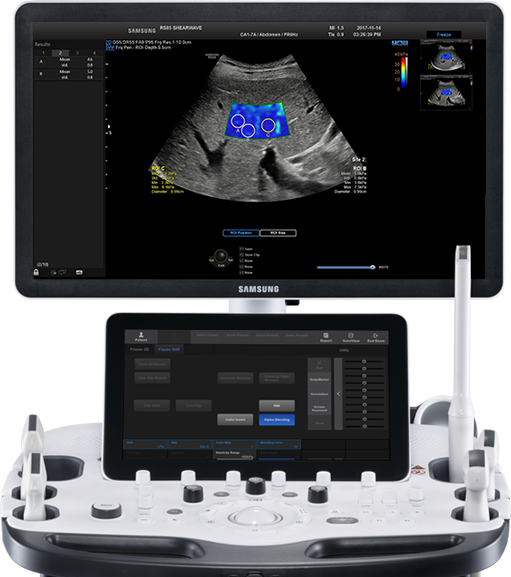
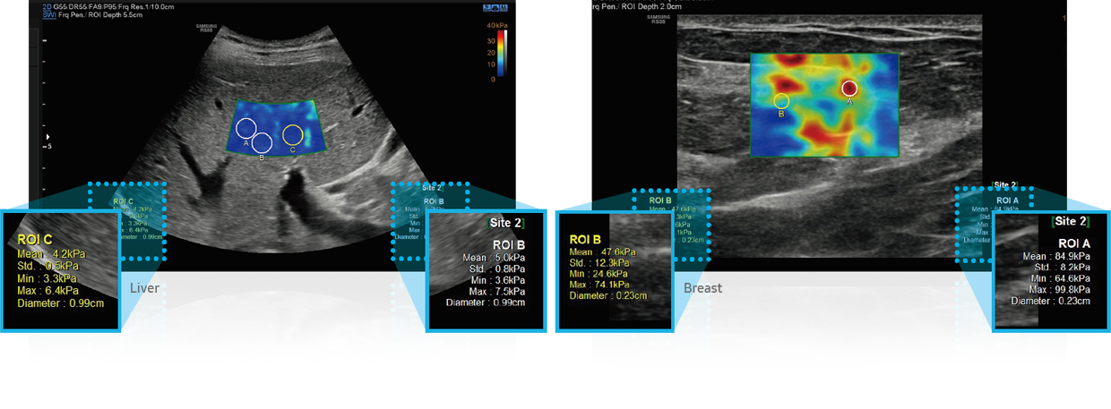
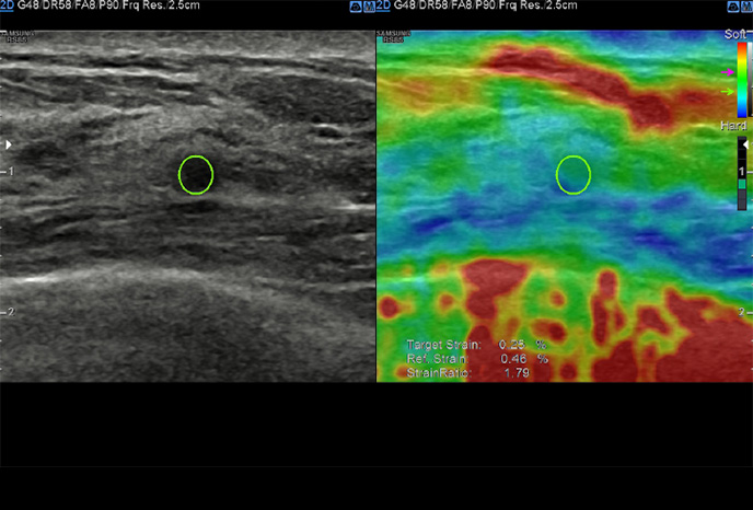
S-Shearwave Imaging™ allows for non-invasive assessment of the stiffness of tissue/lesions in the breast and liver, by providing an advanced level of diagnostic information.
MV-Flow™
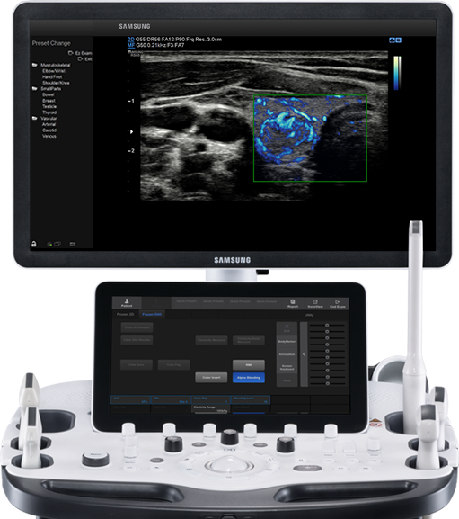
MV-Flow™ offers a novel alternative to Color Doppler for visualizing slow flow microvascularized structures.

Samsung’s premium image-enhancing and artifact-suppressing technologies provide clear, detailed imaging that you can count onto help improve diagnostic confidence and imaging continuity as well as its expert tools offer new perspectives andprovide additional information for confident decision making.
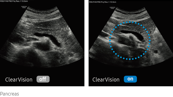
The noise reduction filter improves edge enhancement and creates sharper 2D images for optimal diagnostic performance. The integration of specialized Samsung technology results in a notable improvement in image quality. In addition, ClearVison provides application-specific optimization and advanced temporal resolution in live scan mode.
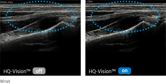
HQ-Vision™ is a new, advanced technology for visualizing anatomical structures. With improved image clarity, this feature helps make a reliable diagnosis quickly.
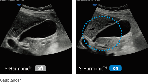
This new harmonic technology improves image clarity, near to far. Reducing signal noise, S-Harmonic™ provides more uniform ultrasound images. Combined with the S-Vue™ transducers, S-Harmonic™ takes RS80 EVO image quality one step further.
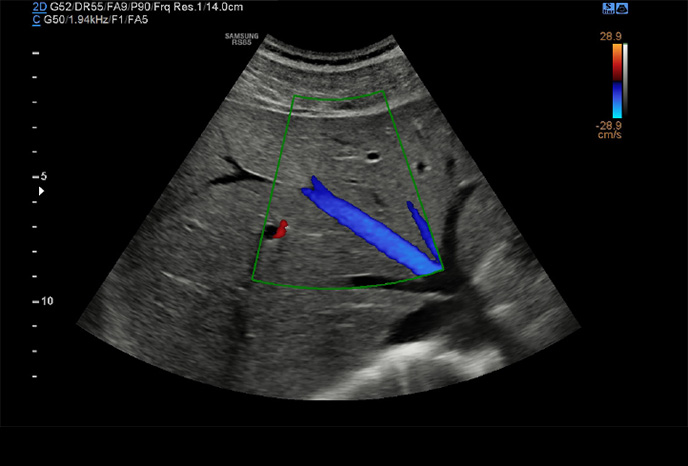
LumiFlow™ is a function that visualizes blood flow in three dimensional-like to help understand the structure of blood flow and small vessels intuitively.
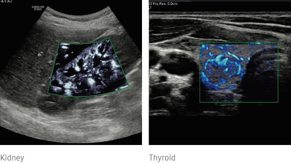
MV-Flow™ offers a novel alternative to Color Doppler for visualizing slow flow microvascularized structures. High frame rates and advanced filtering enable MV-Flow™ to provide a detailed view of blood flow in relation to surrounding tissue or pathology with enhanced spatial resolution and temporal resolution.
Intuitive multi-modality fusion imaging with high precision
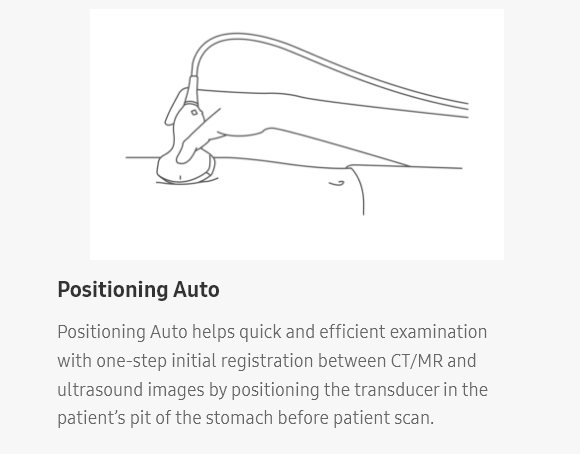
S-Fusion™ enables simultaneous localization of a lesion using real-time ultrasound in conjunction with other volumetric imaging modalities. Samsung’s Auto Registration helps quickly and precisely fuse the images, increasing efficiency and reducing procedure time. S-Fusion™ enables precise targeting during interventional and other advanced clinical procedures.
S-Fusion™ for ProstateAssists in precise targeting during prostate biopsiesS-Fusion™ for Prostate allows safe navigation and precise targeting during prostate biopsies based on 3D models created from MR data sets, and also provides a function to report biopsy location.
Sophisticated 2D & Color Images Processed by CrystalPure™
CrystalPure™ imaging engine help you to make more confident diagnoses with fundamental 2D images and enhanced color performance. It also lessens the incidence of clutter and boosts the level of color signal processing.
CEUS+ technology uses the unique properties of ultrasound contrast agents. When stimulated with low acoustic pressure, the oscillating microbubbles reflect both fundamental and harmonic frequency signals. In addition, Samsung’s technologies provide a clear visualization of vessels and blood flow for a more informed and confident diagnosis.
The function uses directional power doppler technology, enabling you to examine even the peripheral vessels. It displays information on the intensity and direction of blood flow. DPDI Mode: When it is selected, the PRF value is displayed on the color bar.
With its advanced intelligent solutions, including an extensive range of quantification functions,RS80 EVO provides measurement consistency while reducing variability between users.
Non-invasive quantification method of tissue stiffness
S-Shearwave Imaging™ allows for non-invasive assessment of the stiffness of tissue/lesions in the breast and liver,by providing an advanced level of diagnostic information. The color-coded elastogram, quantitative measurements (inkPa or m/s),dual or single display option, and user-selectable ROI (position and size) functions are especially useful for the accurate diagnosis of breast and liver diseases.
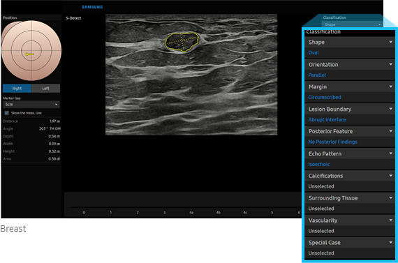
Semi-automated imaging reporting tool for breast assessment
The feature, which analyzes selected lesions in the breast ultrasound study and shows the analysis data, applies BI-RADS ATLAS* (Breast Imaging-Reporting and Data System, Atlas) to provide standardized reporting; and helps diagnosis with the streamlined workflow.
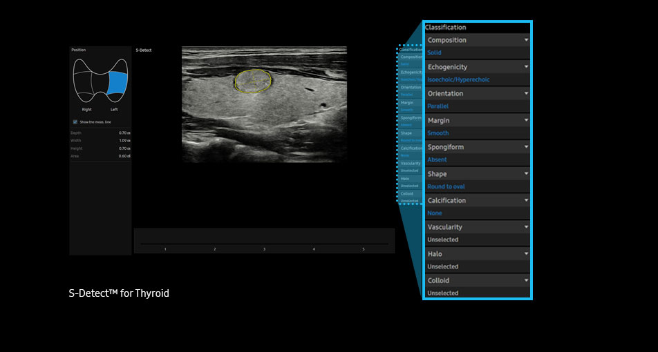
Semi-automated imaging reporting tool for thyroid assessment
The feature, which analyzes selected lesions in the thyroid ultrasound study and shows the analysis data, provides standardized reporting based on the *ATA, BTA, EU-TIRADS, K-TIRADS and ACR TI-RADS guidelines; and helps diagnosis with the streamlined workflow.
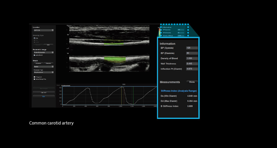
Tool for detecting functional changes of cardiovascular vessels Arterial Analysis™ detects functional changes of vessels, providing measurement values such as the stiffness, intima-media thickness and pulse wave velocity of the common carotid artery. Since the functional changes occur before morphological changes, this technology supports the early detection of cardiovascular disease.
AutoIMT+ is a screening tool to analyze a patient’s potential risk of cardiovascular disease. It allows easy intima-media thickness measurement of both the anterior and posterior wall of the common carotid by the click of a button.
S-3D Arterial Analysis™ simplifies volume measurement of arterial plaque, providing 3D vessel modeling. With Samsung’s S-3D Arterial Analysis™, obtaining information on the arterial plaque volume is surprisingly fast and easy even on difficult patients. In addition, it allows you to track the morphological changes of the artery.
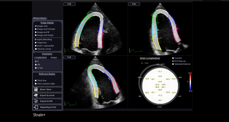
Strain+ is a quantitative tool for measuring global and segmental wall motion of the left ventricle (LV). In Strain+, three standard LV views and a Bull’s Eye are displayed in a quad screen for easy and quick assessment of the LV function.
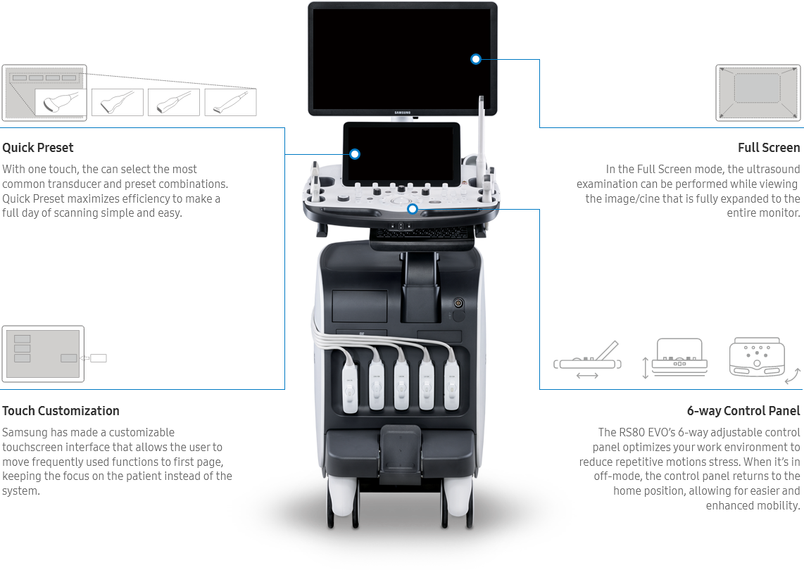
The RS80 EVO has been designed to streamline your workflow by enhancing efficiencythrough reducing keystrokes and by combining multiple actions into one.
Real-time image sharing solution
SonoSync™ is a real-time image sharing solution that allows collaborative communication and remote controllability for effective collaborationbetween physicians and sonographers at different locations. Apart from these, SonoSync™ has several other elegant features like marking, invitation,still image sharing, multi-user, and multi-view. SonoSync™ brings telesonography into reality.


Automatic transducer setting tool based on the worklist EzPrep™ is a function that automatically selects the transducer based on the worklist inputted in the ultrasound system and sets the Preset of the selected transducer

RIS Browser is a function that improves the workflow in the hospital by allowing access to RIS through the browser embedded in the system for the post process without need to move to the PC after scanning.

Tools for protecting against cyber threats from external attacks

Strengthened surveillance for tracking the access of patient information

Encryption functions for safeguarding data whether at-rest or in-transit
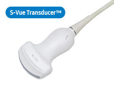
Application: Abdomen, Obstetrics, Gynecology, Musculoskeletal, Pediatric, Vascular, Urology
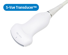
Application: Abdomen, Obstetrics, Gynecology, Musculoskeletal, Pediatric, Vascular, Urology
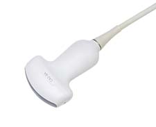
Application: Abdomen, Obstetrics, Gynecology
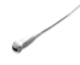
Application: Pediatric, Vascular
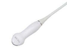
Application: Pediatric, Vascular
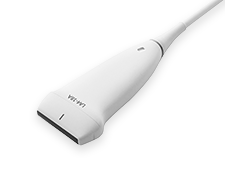
Lorem ipsum dolor sit amet, consectetur adipiscing elit. Ut elit tellus, luctus nec ullamcorper mattis, pulvinar dapibus leo.
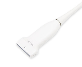
Lorem ipsum dolor sit amet, consectetur adipiscing elit. Ut elit tellus, luctus nec ullamcorper mattis, pulvinar dapibus leo.
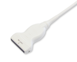
Lorem ipsum dolor sit amet, consectetur adipiscing elit. Ut elit tellus, luctus nec ullamcorper mattis, pulvinar dapibus leo.
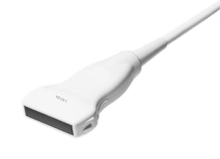
Lorem ipsum dolor sit amet, consectetur adipiscing elit. Ut elit tellus, luctus nec ullamcorper mattis, pulvinar dapibus leo.
Application: Abdomen, Obstetrics, Gynecology
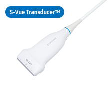
Lorem ipsum dolor sit amet, consectetur adipiscing elit. Ut elit tellus, luctus nec ullamcorper mattis, pulvinar dapibus leo.
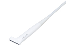
Lorem ipsum dolor sit amet, consectetur adipiscing elit. Ut elit tellus, luctus nec ullamcorper mattis, pulvinar dapibus leo.
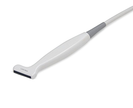
Lorem ipsum dolor sit amet, consectetur adipiscing elit. Ut elit tellus, luctus nec ullamcorper mattis, pulvinar dapibus leo.
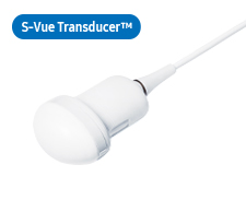
Application: Abdomen, Obstetrics, Gynecology
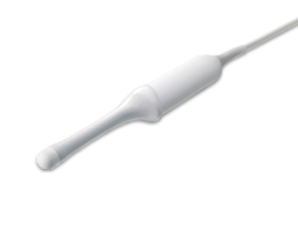
Application: Obstetrics, Gynecology, Urology
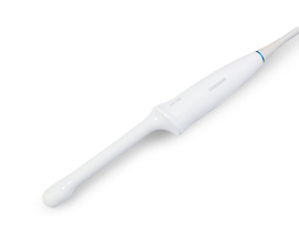
Application: Obstetrics, Gynecology, Urology
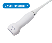
Application: Cardiac, TCD, Abdomen
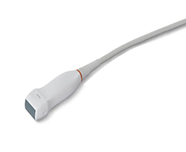
Application: Cardiac, TCD, Abdomen
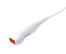
Application: Cardiac, Vascular, Pediatric
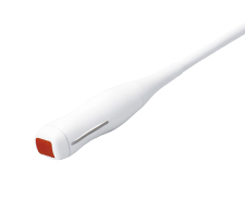
Application: Cardiac, Pediatric
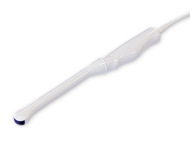
Application: Obstetrics, Gynecology, Urology
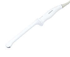
Application: Obstetrics, Gynecology, Urology
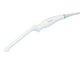
Application: Obstetrics, Gynecology, Urology
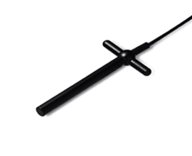
Application: Cardiac, Vascular
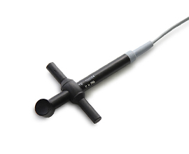
Application: Cardiac
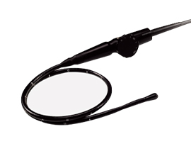
Application: Cardiac
